Leaf is the main place where photosynthesis occurs. Most leaves are usually green, due to presence of chlorophyll in the leaf cells. However, some leaves may have different colors, caused by other plant pigments that mask the green chlorophyll. In this article, learn the difference between monocot and dicot leaves. The basis of comparison include: Stomata, Shape, leaf orientation, Upper and lower surface color, intellectual spaces, Bundle Sheath, Mesophyll differentiation, venation pattern, the hypodermis of the midrib.

Monocot Leaf
Monocot leaves are leaves which appear on plants produced from seeds with single cotyledon like maize, rice, grass, wheat etc. The monocot leaves are usually described as isobilateral leaves because the both the upper and lower surfaces have the same color.
In a monocot leaf equal number of stomata is present on both surfaces of the epidermis. This condition is normally described as amphi stomatic condition.
Anatomy of a monocot leaf
Upper Epidermis
The upper epidermis is a single layer made up of cubical shaped cells with no intercellular spaces in between them. The outer surface of the upper epidermis cell is covered by a thin cuticle. A few cells present in the upper epidermis are enlarged to form motor cells referred to as bulliform cells. These cells help the leaf to roll over themselves in order to reduce the surface area exposed to sunlight during hot seasons.
Mesophyll
Mesophyllis a green tissue between upper epidermis and lower epidermis. In monocot leaf, the mesophyll tissue is not differentiated into palisade parenchyma and spongy parenchyma with chloroplast and chlorophyll. The mesophyll is usually involved in photosynthesis process in the leaves of these plants.
Vascular Bundles
Vascular bundles represent the veins of the leaves. Each vascular bundle consists of phloem and xylem tissues surrounded by a bundle sheath. Bundle sheath layer of the vascular bundle is made up of large barrel shaped endodermal cells. Xylem is usually responsible for conduction of water and dissolved minerals whereas phloem is responsible for conduction of dissolved food materials.
Dicot Leaf
Dicotyledons commonly known as dicots include mango, peanut, peas, oranges, cashews, beans, apples, oak trees etc.
Anatomy of a dicot leaf
Just like a monocot leaf, the main internal structures of a dicot leaf include: epidermis, mesophyll and vascular bundle.
Epidermis
The epidermis is usually made up of a single layer of cells that are closely packed. A dicot leaf consist of a lower and upper epidermis with small openings referred to as stomata. The upper epidermis is thicker than the lower epidermis. More importantly, the lower epidermis has more stomata than the upper epidermis. The main function of the epidermis is to give protection to the inner tissue known as mesophyll.
Mesophyll
The mesophyll usually has two regions the spongy and palisade parenchyma. The palisade parenchyma cells contain more chloroplasts than the spongy parenchyma cells and thus its function is photosynthesis. On the other hand, spongy cells are irregularly shaped and loosely arranged so as to facilitate the exchange of gases within the air spaces.
Vascular bundles
The vascular bundles of a dictot leaf are surrounded by a compact layer of paranchymotous cells known as border parenchyma. The xylem consists of metaxylem vessels and protoxylem vessels. Phloem consists of sieve tubes, companion cells and phloem parenchyma.
The Key differences
- The main characteristic feature that differentiates a monocot and a dicot leaf is that, the guard cells of stomata are kidney-shaped in dicot leaf and dumb-bell shaped in a monocot leaf.
- The orientation of a dicot leaf is dorsiventral while that of a monocot leaf is isobilateral. A dorsiventral organ is one that has two surfaces differing from each other in appearance and structure. Isobilateral orientation is whereby plant leaf surface parts (upper and lower) are identical to each other.
- The upper surface of a dicot leaf is dark green while the lower surface is light green in color. On the other hand, the upper and lower surfaces of a monocot leaf are equally green.
- The vascular bundle is large in dicot leaf whereas in monocot leaf, both small and large vascular bundles are present.
- In a dicot leaf stomata are usually present on the lower surface of the leaf, a condition referred to as hypostomatic. On the contrary, the leaves of monocot plant have stomata on both surface of the leaf, a condition referred to as amphistomatic.
- The intercellular spaces of a dicot plant leaf are relatively large due to presence of loosely packed mesophyll cells. In monocot plant leaf, the intercellular spaces are relatively small due to compact arrangement of mesophyll cells.
- The walls of epidermal cells of a dicot leaf do not have silica deposition whereas; the walls of epidermal cells of a monocot plant leaf have heavy deposition of silica.
- The stomata are arranged randomly on the epidermis of a dicot plant leaf whereas in monocot leaf, the stomata are arranged in parallel rows and are uniformly present on both the leaf surfaces.
- The bundle sheath of a dicot plant leaf generally has a single layer and formed of colorless cells. On the contrary, the bundle sheath of a monocot plant leaf may have a single or double layer and formed of colored cells due to presence of chloroplasts.
- The mesophyll of a dicot leaf is differentiated into two parts, the lower spongy mesophyll and upper palisade. On the contrary, the mesophyll of a monocot plant leaf has no such differentiation.
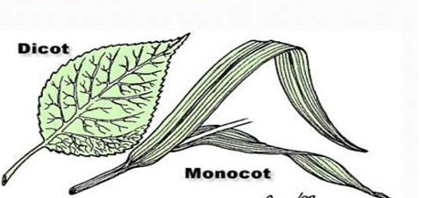
- The venation pattern in a dicot plant leaf is reticulate (veins are interconnected and form a web like network). In contrast, the venation pattern of a monocot plant leaf is parallel (whereby the secondary veins run parallel to each other off a central, perpendicular primary vein).
- The shape of a dicot plant leaf is broader and relatively smaller whereas a monocot plant leaf is slender and long in shape.
- The hypodermis of the midrib region of a dicot plant leaf is collenchymatous while in a monocot plant leaf, the hypodermis of the midrib region is sclerenchymatous.
- In monocot leaf, large vascular bundles may show differentiation into protoxylem and meta-xylem elements whereas; in dicot leaf large vascular bundles do not show differentiation into protoxylem elements.
- The bundle sheath extension of a dicot leaf is parenchymatous whereas the bundle sheath extension of a monocot leaf is sclerenchymatous.
- The bulliform (Motor) cells are absent in the epidermis of a dicot plant leaf. In contrast, the bulliform (motor) cells are very much present in the epidermis of a monocot plant leaf.
Difference Between Dicot And Monocot Leaf In Tabular Form
| THE BASIS OF COMPARISON | DICOT LEAF | MONOCOT LEAF |
| Stomata | The guard cells of stomata are kidney-shaped in dicot leaf | The guard cells of stomata are dumb-bell shaped in monocot leaf. |
| Shape | The shape of a dicot plant leaf is broader and relatively smaller. | A monocot plant leaf is slender and long in shape. |
| Leaf Orientation | The orientation of a dicot leaf is dorsiventral. | The orientation of a monocot leaf is isobilateral. |
| Upper and Lower Surface Color | The upper surface of a dicot leaf is dark green while the lower surface is light green in color. | The upper and lower surfaces of a monocot leaf are equally green. |
| Size of the Vascular Bundles | The vascular bundle is large in dicot leaf. | Both small and large vascular bundles are present. |
| Stomata Arrangement | In a dicot leaf stomata are usually present on the lower surface of the leaf, a condition referred to as hypostomatic. | The leaves of monocot plant have stomata on both surface of the leaf, a condition referred to as amphistomatic. |
| Intercellular Spaces | The intercellular spaces of a dicot plant leaf are relatively large due to presence of loosely packed mesophyll cells. | In monocot plant leaf, the intercellular spaces are relatively small due to compact arrangement of mesophyll cells. |
| Silica Deposition | The walls of epidermal cells of a dicot leaf do not have silica deposition. | The walls of epidermal cells of a monocot plant leaf have heavy deposition of silica. |
| Stomata Arrangement | The stomata are arranged randomly on the epidermis of a dicot plant leaf. | The stomata are arranged in parallel rows and are uniformly present on both the leaf surfaces. |
| Bundle Sheath | The bundle sheath of a dicot plant leaf generally has a single layer and formed of colorless cells. | The bundle sheath of a monocot plant leaf may have a single or double layer and formed of colored cells due to presence of chloroplasts. |
| Mesophyll Differentiation | The mesophyll of a dicot leaf is differentiated into two parts, the lower spongy mesophyll and upper palisade. | The mesophyll of a monocot plant leaf has no such differentiation. |
| Venation Pattern | The venation pattern in a dicot plant leaf is reticulate (veins are interconnected and form a web like network). | The venation pattern of a monocot plant leaf is parallel (whereby the secondary veins run parallel to each other off a central, perpendicular primary vein). |
| The Hypodermis of the Midrib | The hypodermis of the midrib region of a dicot plant leaf is collenchymatous. | In a monocot plant leaf, the hypodermis of the midrib region is sclerenchymatous. |
| Vascular Bundle Differentiation | In dicot leaf large vascular bundles do not show differentiation into protoxylem elements. | In monocot leaf, large vascular bundles may show differentiation into protoxylem and meta-xylem elements. |
| Nature of the Bundle Sheath Extension | The bundle sheath extension of a dicot leaf is parenchymatous | The bundle sheath extension of a monocot leaf is sclerenchymatous. |
| The Bulliform (Motor) Cells | The bulliform (Motor) cells are absent in the epidermis of a dicot plant leaf. | The bulliform (motor) cells are very much present in the epidermis of a monocot plant leaf. |
What are the Similarities Between Monocot and Dicot Leaves
- Both have vascular bundles with a bundle sheath extension.
- Both monocot and dicot leaves are differentiated internally into mesophyll , epidermis and vascular tissues.
- They both possess chloroplasts.
- In monocot and dicot leaves, xylem and phloem consist of protoxylem and protophloem; and metaxylem and metaphloem.
- Both monocot and dicot leaves contain stomata and guard cells.
- Vascular bundles are conjoint and collateral in both monocot and dicot leaves.
- In both monocot and dicot leaves the major portions of the ground tissue is parenchymatous.
- Hypodermis is present in both dicot and monocot leaves.