Monocot Root
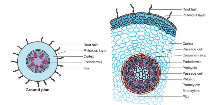
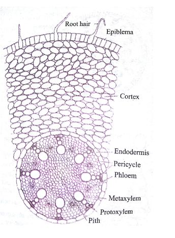
The tissue at the center of monocot roots consists of (vascular bundle) xylem and phloem and it is surrounded by the cortex which is made up of parenchyma cells. The outmost layer of the root is referred to as the epidermis followed by the endodermis or sclerenchyma. The endodermis is an inner layer of cells surrounding the vascular bundle. The endodermis and phloem are separated by a layer of cells referred to as the pericycle where root branching occurs.

Features Of Monocot Roots You Need To Know
- Epiblema or Epidermis– is a single layered, thin walled colorless, polygonal without intercellular spaces, with presence of unicellular root hairs hence referred to as rhizoids or piliferous layer. The root hairs and epidermal cells take part in the absorption of water and minerals from the soil.
- Pericycle: This is the outmost layer of stellar system. it is composed of uniseriate layer of parenchymatous cells without intercellular spaces. Several lateral roots arise from this structure.
- Secondary growth is absent in monocot roots due to lack of vascular and cork cambium.
- Cortex: This is a multi-layered well developed structure, made from oval parenchymatous cells with intercellular spaces. Cortex helps in mechanical support to the roots. Also, Cortex gives rise to exodermis (sclerenchymatous region). Cortex may be heterogeneous with outer dead exodermis.
- Vascular Bundles:are radial, xylem is exarch i.e the Protoxylem lies towards periphery and Metaxylem towards the center. Bundles more than six. Metaxylem elements are oval or circular. The phloem is also exarch (protophloem towards the periphery and Metaphloem towards the center). The number of xylem and phloem vary from 8 to 46 (100 in pandanus). Phloem fibers are absent.
- Conjunctive tissues arelimited or even absent. Conjunctive tissue is a parenchymatous tissue which separate xylem and phloem tissues.
- Endodermis:This is the innermost layer of cortex made from barrel shaped parenchyma. The endodermis is characterized by presence of casparian stripes.
- Passage Cells:usually passage cells are absent in monocot roots.
- Pith is large, well developed portion of monocot root. It contains abundant amount of starch grains.
Dicot Root
A carrot is usually a perfect example of dicot root. The vascular bundles of dicot roots are arranged in the form of one or two broken rings. These bundles are definite in shape, size and are smaller as compared to the bundles in monocot rots. Dicot roots normally have their xylem in the center of the root and phloem, outside the xylem.

Features Of Dicot Roots You Need To Know
- Epiblema or Epidermis– it is made up of thin walled, compactly arranged living parenchymatous cells. Epidermis/Epiblema is characterized by absence of stomata and cuticle. It provides protection to the roots due to presence of unicellular root hair.
- Pericycle. Some dicots do not bear pericycle. Pericycle becomes meristematic to give secondary roots and secondary tissues.
- Secondary growth is present in dicot roots due to presence of vascular and cork cambium.
- Vascular bundles. They are between 2 to 8 in number, radial and arranged in ring. Xylem is exarch, number of xylem varies from 2 to 4. Metaxylems are angular arranged in linear. Phloem fibers are absent or completely reduced.
- Conjunctive tissues are well developed. Conjunctive tissue separate xylem and phloem tissues.
- Pith is very small or completely obliterated. Pith helps in storage of food materials.
- Endodermis consists of barrel shaped compact parenchymatous cells. It contains both casparian stripes and passage cells.
- Cortex is homogenous i.e without any differentiation.
- Passage Cells:usually passage cells are present in dicot roots.
Also Read: Difference Between Monocot Leaf And Dicot Leaf
Difference Between Dicot And Monocot Root In Tabular Form
| BASIS OF COMPARISON | MONOCOT ROOT | DICOT ROOT |
| Cortex | Cortex may be heterogeneous (differentiated) with outer dead exodermis. | Cortex is homogenous i.e without any differentiation. |
| Pericycle | Pericycle in monocot roots, only produces the lateral roots. | The pericycle gives rise to lateral roots, cork cambium and the part of the vascular cambium. |
| Pith | Pith is large, well developed portion of monocot root. It contains abundant amount of starch grains. | Pith is very small or completely obliterated. |
| Xylem &Phloem | Xylem and phloem are numerous in number. | Xylem and phloem are limited in numbers. |
| Passage Cells | Usually passage cells are absent in endodermis of monocot roots. | Usually passage cells are present in endodermis of dicot roots. |
| Secondary Growth | Secondarygrowth is absent in monocot roots due to lack of vascular and cork cambium. | Secondarygrowth is present in dicot roots due to presence of vascular and cork cambium. |
| Phloem fibers/phloem sclerenchyma | Phloem fibers are completely absent. | Phloem fibers are completely reduced. |
| Casparian Strips | Casparian strips are very much present in the endodermis, they help the endodermis to form water tight jacket around the vascular tissues. | Some endodermal cells near Protoxylem have no casparian strips. |
| Conjunctive Tissues | Conjunctivetissuesarelimited or even absent. | Conjunctivetissues are well developed. |
| Metaxylem | Metaxylem elements are oval or circular. | Metaxylem elements are angular, arranged in linear position. |
What Are Some of Similarities Between Monocot And Dicot Roots?
- In monocot and dicot roots, xylem is exarch i.e protoxylem lies towards the periphery and Metaxylem towards the center.
- In both dicot and monocot roots, endodermis consists of barrel shaped parenchyma without intercellular spaces.
- In both monocot and dicot roots, pith consist of thin walled, polygonal parenchyma cells with intercellular spaces.
- Both monocot and dicot roots have radial vascular bundles.