Gram Negative Bacteria & Gram Positive Bacteria
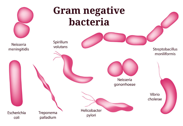
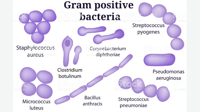
The “gram-positive” and “gram negative” are terms used by microbiologists to classify bacteria. The classification is based on the bacterium’s chemical and physical cell wall properties. For proper determination as to weather a bacteria is gram-positive or gram-negative, microbiologist usually performs a special type of staining technique referred to as Gram Stain. The name gram stain comes from the name of the person who discovered it, Han Christian Gram (1884). The staining method uses crystal violet dye which is retained by the peptidoglycan cell wall of both gram negative and gram positive bacteria. This stain will either stain the cells purple (for positive) or pink (for negative).
What You Need To know About Gram Negative Bacteria (Major Characteristics)

- These are bacteria that are not able to retain the crystal violet color and show negative result to gram stain test.
- With exception of Neisseria, the rest are usually non-spore forming rods.
- The thickness of the cell wall is between 8 nm and 12 nm.
- More elastic and less rigid. The elasticity of the cell wall is due to the less amount of peptidoglycan, which is between 2-12%.
- The cell wall is sensitive to alkali.
- The cell wall is less sensitive to degradation by action of the lysozyme.
- Most pathogenic bacteria belong to gram negative group.
- There is several variety of amino acid in the cell wall of gram negative.
- Usually not found to produce endospores.
- Proteinaceous membrane channel referred to as porins are present.
- Lipid content is high. It is usually between 11-14%.
- The ration of RNA to DNA is 1:1
What You Need To Know About Gram Positive Bacteria (Major Characteristics)

- These are bacteria that give positive result to the gram stain test and take up the crystal violet stain.
- With exception of Lactobacillus and Corynebacterium, the rest are usually spore forming rods.
- The thickness of the cell wall can be up to 15-30 nm or sometimes can be 80 nm.
- The cell wall shows some resistance to alkali.
- Less elastic and more rigid. The rigidity of the cell wall is due to the high amount of peptodoglycan, which is between 70-80%.
- Cell wall contains muramic acid, which is between 16-20% of the total dry weight.
- There is few variety of amino acid in the cell wall.
- Cell wall contains muramic acid, which is between 16-20% of the total dry weight.
- The cell wall is very much susceptible to degradation by the action of the lysonzyme.
- Some produce endospores during unfavorable conditions.
- Few pathogenic bacteria belong to gram positive group.
- The flagellar structure has two rings in the basal body.
- Lipid content is low. It is usually between 1-4%.
- The ratio of RNA to DNA is 8:1
Also Read: Difference Between Staphylococcus And Streptococcus Bacteria
Difference Between Gram Positive And Gram Negative Bacteria In Tabular Form
| BASIS OF COMPARISON | GRAM NEGATIVE BACTERIA | GRAM POSITIVE BACTERIA |
| Description | These are bacteria that are not able to retain the crystal violet color and show negative result to gram stain test. | These are bacteria that give positive result to the gram stain test and take up the crystal violet stain. |
| Washing With De-staining Solution | Color of crystal violet will not be retained after washing with de-staining solution (alcohol). | Retain the color of crystal violet after washing with alcohol (de-staining). |
| Morphology | With exception of Neisseria, the rest are usually non-spore forming rods. | With exception of Lactobacillus and Corynebacterium, the rest are usually spore forming rods. |
| Cell Wall Thickness | The thickness of the cell wall is between 8 nm and 12 nm. | The thickness of the cell wall can be up to 15-30 nm or sometimes can be 80 nm. |
| Other Features of Cell Wall | The cell wall is sensitive to alkali. | The cell wall shows some resistance to alkali. |
| More elastic and less rigid. | Less elastic and more rigid. | |
| The elasticity of the cell wall is due to the less amount of peptidoglycan, which is between 2-12%. | The rigidity of the cell wall is due to the high amount of peptodoglycan, which is between 70-80%. | |
| Bilayered. | Single layered. | |
| The content of muramic acid is between 2-5% of the total dry weight. | Cell wall contains muramic acid, which is between 16-20% of the total dry weight. | |
| Teichoic acid is absent in the cell wall. | Teichoic acid is very much present in the cell wall. | |
| Cell wall is sensitive to alkalis and soluble in 1% KOH solution. | Cell wall is resistant to alkalis and insoluble in 1% KOH solution. | |
| There is several variety of amino acid in the cell wall. | There is few variety of amino acid in the cell wall. | |
| The cell wall is less sensitive to degradation by action of the lysozyme. | The cell wall is very much susceptible to degradation by the action of the lysonzyme. | |
| Wavy and uneven | Straight and even. | |
| Aromatic and sulfur-containing amino acid in the cell wall is present. | Aromatic and sulfur-containing amino acid in the cell wall is absent. | |
| S-layer is attached to the outer membrane. | S-layer is attached to the peptidoglycan layer. | |
| Examples | Escherichia coli Salmonella Klebsiella Proteus Helicobacter Pseudomonas Vibrio Rhizobium Acetobacter | Staphylococcus Streptococcus Bacillus Clostridium Enterococcus |
| Endospore Formation | Usually not found to produce endospores. | Some produce endospores during unfavorable conditions. |
| Gram Staining Reaction | Accept safranin after decolorization and stain pink or red on Gram’s staining. | Retains crystal violet dye and stain blue or purple on Gram’s staining. |
| Pathogenicity | Most pathogenic bacteria belong to gram negative group. | Few pathogenic bacteria belong to gram positive group. |
| Toxins Produced | Endotoxins or Exotoxins | Exotoxins |
| Porins | Proteinaceous membrane channel referred to as porins are present. | Proteinaceous membrane channel referred to as porins are absent. |
| Flagellar Structure | The flagellar structure contains four rings in the basal body. | The flagellar structure has two rings in the basal body. |
| Lipopolysaccharides | Present | Absent |
| Periplasmic space | Periplasmic space is present. | Periplasmic space is absent and if present, then, it is very narrow. |
| Lipid Content | Lipid content is high. It is usually between 11-14%. | Lipid content is low. It is usually between 1-4%. |
| Outer Membrane | The outer membrane is present. | The outer membrane is absent. |
| Ratio Of RNA:DNA | The ration of RNA to DNA is 1:1 | The ratio of RNA to DNA is 8:1 |
| Sodium Azide | Show low resistance to sodium azide solution. | Show high resistance to sodium azide solution. |
| Mesosomes | Less prominent. | Very much prominent |
| Magnetosomes | Present | Usually absent. |
| Resistance To Drying | Low | High |
| Inhibition By Basic Dyes | Low | High |
| Resistance To Physical Disruption | Low | High |
| Resistance To Sodium Azide | Low | High |
| Susceptibility To Anionic Detergents | Low | High |
| Susceptibility To Penicillin And Sulfonamide | Low | High |
| Susceptibility To Streptomycin, Chloramphenicol And Tetracycline | High | Low |
Also Read: Difference Between Bacteria And Virus
What Are Some of the similarities Between Gram Positive Bacteria And Gram Negative Bacteria?
- Both gram positive and gram negative bacteria contain extra-chromosomal genetic materials (Plasmids).
- They both have a surface layer referred to as an S-layer.
- Both are bacterial cells and contain many flagellated and non-flagellated species.
- Both gram negative and gram positive bacteria have covalently closed circular DNA as the genetic material.
- Both gram positive and gram negative bacteria undergo binary fission as a mode of asexual reproduction.
- Gram negative and gram positive bacteria, both lack membrane bound organelles.
- Both are prokaryotic in nature.
- In both groups, cell wall is made up of peptidoglycan.
- Gram positive and gram negative bacteria undergo genetic recombination through transduction, transformation and conjugation.
- In both groups, cytoplasm is surrounded by lipid bilayer with many membrane spanning proteins.