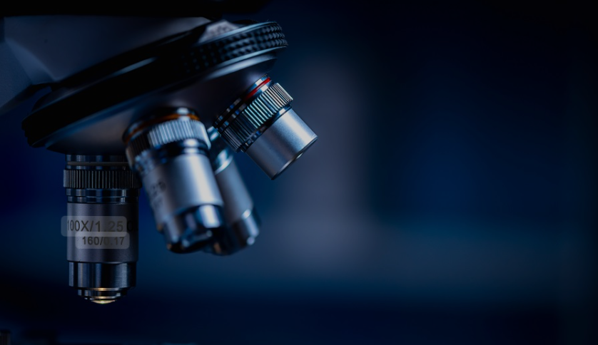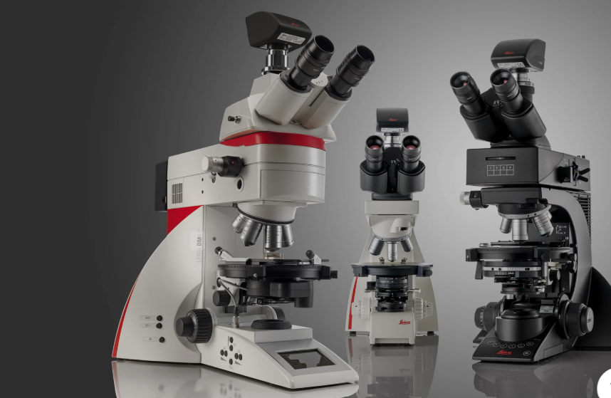
A microscope is a scientific instrument used to magnify and observe objects that are too small to be seen with the naked eye. It works by using lenses, light, or other forms of energy such as electrons or sound waves to enlarge the image of a specimen. Microscopes have become essential tools in science, medicine, and industry, allowing researchers to explore the intricate details of cells, microorganisms, and even atomic structures.
The invention of the microscope marked a turning point in scientific discovery. Early optical microscopes used simple glass lenses to magnify samples, leading to the first observations of bacteria, cells, and tissues. Over time, technological advancements gave rise to more powerful and specialized microscopes, including electron and digital types. These developments opened new frontiers in biology, physics, and materials science.
Microscopes function based on various principles depending on their design. Optical microscopes use visible light and glass lenses, while electron microscopes use beams of electrons to achieve far higher magnifications. Other types, like atomic force or scanning tunneling microscopes, rely on physical interactions rather than light. Each type serves a distinct purpose, from studying living cells to analyzing nanomaterials or industrial defects.
The importance of microscopes extends beyond laboratories. In medicine, they are used to diagnose diseases by examining tissues and microorganisms. In education, they help students understand the microscopic world. In fields like geology, engineering, and forensics, microscopes assist in analyzing materials, identifying substances, and ensuring product quality. Their versatility makes them invaluable across countless disciplines.
Modern microscopes now incorporate digital imaging and computer software, allowing scientists to capture, analyze, and share microscopic data easily. Techniques such as fluorescence and confocal microscopy enable the visualization of complex biological processes in real time. This integration of technology continues to enhance precision, efficiency, and accessibility in scientific research.

Types of Microscopes
Compound Microscope
The compound microscope is one of the most common types used in laboratories and classrooms. It uses two sets of lenses — the objective and the eyepiece — to magnify small specimens like cells, bacteria, and tissues.
This microscope relies on transmitted light to illuminate thin samples, producing a clear, high-resolution image. It can magnify objects up to around 1,000 times, making it ideal for biological and medical studies.
Stereo Microscope (Dissecting Microscope)
A stereo microscope provides a three-dimensional view of the specimen, making it useful for examining larger or opaque samples. It operates at lower magnifications, typically between 10x and 100x.
Scientists and hobbyists use it for tasks that require precision, such as dissection, circuit board inspection, or studying insects and plants. Its binocular setup gives depth perception that other microscopes lack.
Digital Microscope
Digital microscopes use cameras and computer software instead of eyepieces to display magnified images on a monitor. They often include image capture and measurement capabilities.
These microscopes are popular in research, education, and industry for documentation and analysis. Their digital features make sharing and storing visual data much easier.
Electron Microscope
Electron microscopes use beams of electrons instead of light, allowing them to achieve magnifications up to millions of times. This enables scientists to observe structures at the molecular and atomic level.
They are essential for nanotechnology, materials science, and microbiology research. However, they are expensive and require special conditions such as a vacuum chamber and expert operation.
Transmission Electron Microscope (TEM)
The TEM is a subtype of electron microscope that transmits electrons through ultra-thin specimens. It produces highly detailed, two-dimensional images of internal cell structures or material lattices.
This type of microscope is vital in studying viruses, organelles, and fine material compositions. It provides unparalleled resolution but requires complex sample preparation.
Scanning Electron Microscope (SEM)
The SEM scans a specimen’s surface with focused electron beams to produce a 3D-like image. It shows the texture, shape, and composition of objects with great clarity.
SEM is commonly used in materials science, forensics, and biology to study surfaces and topographies. Its depth of field and detail make it a preferred tool for examining microstructures.
Fluorescence Microscope
Fluorescence microscopes use high-intensity light to excite fluorescent dyes or proteins within samples. The emitted light produces vivid, color-coded images that highlight specific structures.
They are widely used in molecular biology and medical diagnostics to visualize cells, DNA, or proteins. This technology has revolutionized cell imaging and live-cell observation.
Confocal Microscope
Confocal microscopes employ laser light and optical filtering to create high-resolution, 3D images of specimens. They eliminate out-of-focus light, improving clarity and contrast.
Used extensively in biological and biomedical research, confocal microscopy allows scientists to study living tissues and cell dynamics in real time. Its precision enables detailed spatial analysis.
Polarizing Microscope
Polarizing microscopes use polarized light to study materials that affect light differently depending on their orientation. This helps identify minerals, crystals, and fibers.
They are essential tools in geology, chemistry, and forensic science. By analyzing light interference patterns, researchers can determine the composition and structure of various materials.
Phase Contrast Microscope
A phase contrast microscope enhances the contrast of transparent specimens without the need for staining. It converts phase shifts in light into variations in brightness.
This feature makes it ideal for observing live cells, microorganisms, and thin tissues. It allows scientists to study living organisms in their natural state without damaging them.
Scanning Probe Microscope (SPM)
SPMs work by moving a physical probe very close to the specimen’s surface to detect its contours and properties. They can measure electrical, magnetic, and topographical characteristics.
They are highly useful in nanotechnology and surface science, providing detailed 3D maps of surfaces at the atomic scale. Techniques like AFM (Atomic Force Microscopy) fall under this category.
X-Ray Microscope
X-ray microscopes use soft X-rays to penetrate specimens, revealing internal structures without slicing them. They bridge the gap between optical and electron microscopes in terms of resolution.
These microscopes are used in medical imaging, materials research, and nanotechnology. They allow non-destructive imaging of dense or thick samples with high detail.
Atomic Force Microscope (AFM)
The atomic force microscope uses a sharp probe that scans the surface of a sample to measure forces at the atomic level. It does not rely on light or electrons but instead detects interactions between the probe and the surface atoms.
AFM is essential in nanotechnology, materials science, and molecular biology. It provides incredibly detailed 3D surface maps and can even manipulate individual atoms on a material’s surface.
Ultraviolet (UV) Microscope
Ultraviolet microscopes use ultraviolet light instead of visible light to enhance image resolution beyond the limits of traditional optical microscopes. Since UV wavelengths are shorter, they can reveal finer details.
They are mainly used in biological and chemical laboratories to study cellular components and thin materials. However, UV light requires special optics and protection due to its intensity and potential to damage samples.
Infrared (IR) Microscope
Infrared microscopes use infrared radiation to analyze the chemical composition and structure of materials. They are especially effective in studying heat-sensitive or opaque samples.
These microscopes are commonly used in forensic science, material analysis, and semiconductor research. They can detect molecular bonds and provide insights into the chemical makeup of complex specimens.
Acoustic Microscope
Acoustic microscopes use sound waves rather than light or electrons to create detailed images of the interior of materials. They measure variations in acoustic impedance to map internal features.
They are widely used in materials testing, electronics, and medical diagnostics to detect cracks, voids, or hidden defects. This non-destructive technique is valuable for examining delicate or layered structures.
Scanning Tunneling Microscope (STM)
The scanning tunneling microscope works by measuring the flow of electrons (tunneling current) between a sharp metal tip and a conductive surface. It produces atomic-level images of conductive materials.
STM is one of the cornerstones of nanotechnology research, capable of both imaging and manipulating atoms. It provides real-time visualization of electron density and surface structures.
Dark Field Microscope
A dark field microscope enhances contrast by blocking direct light and only capturing scattered light from the specimen. This creates a bright image on a dark background.
It is particularly useful for viewing live or unstained samples, such as bacteria or transparent cells. Researchers use it when they need to observe delicate details that are invisible under normal light.
Petrographic Microscope
Petrographic microscopes are designed specifically for studying thin sections of rocks and minerals under polarized light. They can reveal the mineral composition and texture of geological samples.
These microscopes are essential tools in geology, mining, and material sciences. By analyzing birefringence and crystal structures, scientists can identify mineral types and their formation processes.
Simple Microscope
The simple microscope uses a single lens to magnify small objects. It is the earliest form of microscope and serves as the foundation for more advanced optical systems.
Though basic, it remains useful for educational purposes, quick inspections, and fieldwork. It offers limited magnification but helps demonstrate fundamental optical principles.
Confocal Laser Scanning Microscope (CLSM)
This advanced microscope uses focused laser beams to scan specimens layer by layer, creating sharp 3D reconstructions. It eliminates background blur and allows precise imaging of complex biological samples.
CLSM is used in biomedical research, neuroscience, and cell biology to study structures deep within tissues. Its combination of fluorescence and laser technology provides exceptional resolution and depth clarity.